Safety is our paramount concern at ToNI. Here is a summary of policies we have in place to ensure the safety of everyone who enters our MRI facility.
Zoning
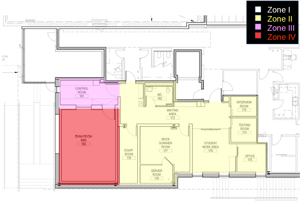
Due to the powerful magnetic field used by the MRI Scanner, access to the MR suite is restricted by establishing the following four zones around the scanner.
- Zone I: Includes all areas that are freely accessible to the general public, and are located outside the MR environment.
- Zone II: This area is the interface between publicly accessible Zone I and the strictly controlled Zones III and IV. Participants are greeted and screened in this zone and are not free to move through Zone II. Participants should be under supervision of MR personnel at all times in this zone.
- Zone III: This area is also known as the Control Room. Access to Zone III is restricted to individuals who have undergone safety screening (users and participants). In this zone, non-MR personnel must be accompanied by, or under the immediate supervision of Level 2 Operators at all times. Additionally, bringing ferromagnetic objects or equipment into this zone should be restricted to essential items, each of which should be monitored by MR personnel.
- Zone IV: This area is also known as the Exam Room. MR Operators should directly monitor the entrance to Zone IV at all times. It is potentially hazardous due to the presence of very strong magnetic fields, as is indicated on the door. Access to Zone IV is restricted to individuals who have undergone safety screening (personnel and participants). In this zone, non-MR personnel must be accompanied by, or under the immediate supervision of Level 2 Operators at all times. No unauthorized objects or equipment should be brought into this area.
Click to Expand

Access and Supervision
- Non-MR personnel must be accompanied by, or under the immediate supervision of, Level 2 Operators for the entirety of their stay within Zones III and IV.
- Non-MR personnel must undergo safety screening (see Safety Screening) before entering Zone III.
- The MR operators should directly observe and control the entrance to Zone IV.
- A lighted sign stating that the “Magnet is Always On” is installed above the entrance to Zone IV.
MRI Operation
- Two MRI Personnel must be present during an MRI session: (1) A Level 2 Operator who runs the MRI scanner and positions the participant and (2) A Level 2 Operator or a Level 1 User who assists the Operator.
- Metal objects are not permitted in the Exam Room as a rule. Any equipment must be examined and authorized by the MR Physicist or Technologist before it is permitted in the Exam Room.
- Participants are always visible to the MRI Operator through a window in the Control Room.
- Participants can hear the MRI Operator through use of the intercom and can exit the scanner at any time via a squeeze ball that alerts the MRI Operator to assist the participant in removal from the machine.
- Noise levels from the MRI scanner can be high. Therefore, participants are given earplugs to reduce MRI noise.
MRI Safety Screening
Screening for participants and non-MR personnel:
- All non-MR personnel wishing to enter Zone III must pass an MR screening process.
- Screening participants should start during the scheduling process, before the participant arrives for a scan. Catch obvious items, such as recent surgical events, implanted metal, and so on, as early in the screening process as possible, well before the day of the scan.
- On the day of the scan, subjects should be screened in a private area (interview room) to make it more likely that the participant discloses their medical history.
- The screening process includes completing a written MR screening questionnaire, as well as orally reviewing the questionnaire with a Level 2 Operator. Each question must be answered.
- The participant (or his/her guardian) and the screening MR personnel must both sign the completed form.
- Any individual undergoing an MR procedure must remove all removable metallic personal belongings and devices on or in them, such as watches, jewelry, cell phones, body piercings, contraceptive diaphragms, metallic drug delivery patches, cosmetics containing metallic particles (e.g., eye make-up), and clothing items that may contain metallic fasteners, hooks, or metallic threads.
- You may wish to screen a subject for metal using a hand-held (“wand” style) metal detector located in the cabinet at the control room. While this can be a useful process, DO NOT substitute a metal detector screen for extensive questioning. Treat it as an optional extra.
- All participants with a history of potential ferromagnetic foreign object penetration must undergo further investigation (e.g., participant’s history, x-ray films, prior CT or MR studies, or documentation about the type of implant or foreign object).
- For participants with implants in their body, the MR compatibility and safety of the implant should be identified using written documents containing information about MR safety testing of the specific make, model, and type of the object. Such information may be provided by the participant, or may be found at www.mrisafety.com
- People with surgical implants in critical organs or in soft tissue (brain, spinal cord, eyes, heart, lungs), must not be allowed to enter the magnet room (Zone IV) regardless of whether the metallic implant/object is magnetic or not. Even when the implanted object is non-magnetic, there is a risk of movement of the object through surrounding soft tissue, causing internal bleeding or other damage.
- It is possible that during an MR imaging examination a ferromagnetic implant or foreign object is discovered within a participant. According to Lenz’s law, rapid motion of the implant perpendicular to the magnetic field can result in forces on the implant that would oppose the motion and may likely be detected by the patient. In such cases, it is important to note that the forces on the implant may increase due to sudden movement. Therefore, the participant should be extricated from the bore with a slow and deliberate rate.
- All non-MR personnel and participants with an implanted cardiac pacemaker, cardioverter defibrillator (ICD), electromechanically activated devices or other electrically conductive devices should be precluded from Zone IV and physically restrained from the 5-Gauss line.
Screening for MR personnel:
- All Level 1 and Level 2 MR personnel are required to go through the screening process.
- All MR personnel must immediately report to the MR director any trauma, procedure, or surgery they undergo where a ferromagnetic object may have become introduced within or on them.
Safety Exclusions
| Conditions That Rule Out a Participant | Conditions That Might Rule Out a Participant | Other Screening Considerations |
|---|---|---|
| Cardiac Pacemaker | Ear Implants | Claustrophobia |
| Surgical Aneurysm Clips | Metal Rods, Plates, or Screws | Physical Discomfort |
| Neurostimulator | Injury to Eyes Involving Metal | Movement Disorders |
| Implanted Pumps | Previous Surgery | Vision/Hearing Problems |
| Metal Fragments in Body/Eyes | IUD | Problems Using Response Devices |
| Pregnancy | Hearing Aids | |
| Nitroglycerin Patch | Dentures | |
| Certain Cochlear Implants | Prosthetic Heart Valve | |
| Weight > 440lbs | Braces | |
| Hair Extensions | ||
| Tattoos or Permanent Eyeliner | ||
| Nicotine Patches |
Things Not to Bring or Wear in the Scanner
- ANYTHING in your pockets
- Metal jewelry (face and body piercing items should be removed)
- Watches
- Hair accessories
- Eye glasses
- Metal on or in clothing (metal buttons, snaps or trimming, underwire bras, belt buckles, metallic fibers)
- Eye shadow (many contain metallic specks that can heat up)
Device and Object Screening
If you are uncertain if a particular device or implant is MR safe, check the list of magnet compatible and incompatible products at www.mrisafety.com. DO NOT scan a participant with an implant not listed as MR safe or cleared as MR conditional for our site’s magnet.

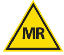
MR Safe
MR Safe indicates no known hazards in all MRI environments. This classification can only be applied to products that do not contain any metallic elements. These products can be considered completely safe with no potential for interaction with the MR field. These items should be identified with a square green “MR-Safe” label.
MR Conditional
These are products that may be brought into certain MRI environments that meet the specified conditions of the “MR Conditional” label for the product. It should be noted that the MR environment in which a device was tested could vary greatly. All “MR Conditional” claims apply to specifically tested static field and gradient field strengths, and only to the precise model, make, and identification of the tested object. Items with an “MR Conditional” rating should be affixed with a triangular yellow “MR Conditional” label.
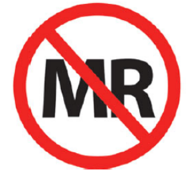
MR Unsafe
MR-Unsafe indicates that an item poses hazards in all MRI environments. These are products that must not be used in an MRI environment. These items should be identified with a round red “MR-Unsafe” label.
External devices which are ferromagnetic and “MR Unsafe” may still, under direct supervision of MR personnel who are familiar with the device and its function, be brought into Zone III. The device must be physically secured or restricted at all times to make sure that they do not inadvertently come too close to the MR scanner.
As a point of safety, all objects should be considered to be magnetic unless they can be positively determined to be non-magnetic (e.g., 100% plastic) or are specially designed to be “MR Safe”. If in doubt, keep it out!
MRI Hazards
All MR Personnel must learn the potential hazards in an MRI facility. They must be able to discuss these hazards with participants, so that potential participants are well-informed before consenting. Additionally, Operators are responsible for considering all potential hazards during a session, whereas, Level 1 Users must not introduce any hazards and must be prepared to assist operators if needed.
Metal objects: When a magnetic object encounters a magnetic field it will experience two types of motion. First, it will try to orient itself such that its longest magnetic axis is parallel to the field direction. Second, the object will tend to move towards the high field region, i.e. it will move towards the center of the magnet. Ferromagnetic metal objects can become projectiles when placed in a strong magnetic field. The strength of the field increases super-linearly with proximity to the magnet bore. Remember, even when you are not scanning, the magnet is on. NEVER bring any metal objects into the scanning rooms.
Auditory considerations: Current passing through the gradient coils during image acquisition produces acoustic noise. Prolonged exposure to this noise may damage the unprotected ear. Hearing protection is required for all participants to reduce the scanner sound pressure to below 99 dB. Also, researchers or parents accompanying the subject must wear earplugs if they are in the magnet room during scanning.
Peripheral nerve stimulation (PNS): Transient application of magnetic field gradients can induce current in conductive materials, including biological tissue. The induced current is greater in peripheral tissue because the amplitude of the gradient is highest farther away from the magnet’s isocenter. Mild skin sensations and involuntary muscle contractions have been reported during scans with fast gradient switching, including echoplanar imaging (EPI). Simply repositioning the subject in the scanner can usually alleviate PNS. To minimize the possibility of PNS, you should instruct your subjects to lie in the magnet with their hands by their sides and with their feet uncrossed. This minimizes large current loops around the body.
Thermal issues: Electrical voltages and currents can be induced within electrically conductive materials that are within the bore of MRI scanner during the MRI imaging process. This might result in the heating of this material by resistive losses. Therefore, all unnecessary or unused electrically conductive materials should be removed from the MR system. Moreover, care should be taken to ensure that no electrically conducting loops (including patient tissue) are formed within the MR scanner during imaging. When electrically conductive materials are required to be within the MRI bore, care should be taken to place thermal insulation between the participant and electrically conductive material. In addition, it is recommended that participant’s tissues do not directly come into contact with the inner bore of the MR scanner during the MRI process. Moreover, the participant’s arms and legs should not form a large loop. Heating of tattooed tissue has been reported, especially with use of iron oxide containing inks. Participants should be informed of the potential for heating or burns and instructed to alert the Operator immediately if warming occurs. For subjects with extensive or dark tattoos, it is recommended that cold compresses or ice packs be placed on the tattooed area throughout the MRI process if these tattoos are within the volume in which the body coil is being used for RF transmission. Additionally, participants with tattoos that had been placed within 48 hours before the MR examination should be advised of the potential for smearing of the edges of the tattoo. There have been rare reports of thermal injuries with clothing that contained electrically conductive material, such as metallic threads and silver impregnated clothing.
Specific Absorption Rate (SAR) issues: Absorption of RF energy by the participant’s body can potentially cause heating of the tissue. Absorption of RF power by the tissue is described in terms of Specific Absorption Rate (SAR). SAR in MRI is a function of many variables, including pulse sequence, coil parameters, the weight of the region exposed, and a participant’s thermoregulatory system (e.g., tissue perfusion, etc.). The scanner automatically estimates SAR level for each pulse sequence based on the participant’s weight and height. Therefore, it is crucial to accurately register the participant’s weight and height in the MRI system.
Pregnancy: Present data have not conclusively documented any deleterious effect of MRI imaging on the fetus. However, given the high susceptibility of the developing fetus to damage in general, it is not worth the risk for pregnant women to participate as subjects in MR research studies. Pregnant MR personnel can work in and around the MR environment throughout all stages of their pregnancy. But, it is recommended not to remain within Zone IV during data acquisition. Pregnant researchers should regulate their own exposure to the magnets.
Pediatric: Children may not be reliable historians and should be screened both in the presence of parents or guardians and separately to maximize the possibility that all potential dangers are disclosed. Pillows, stuffed animals, and other comfort items brought from home should be discouraged from entering Zone IV. Although any age participant may request that others accompany them for their MR examination, this is more common in the pediatric population. Those accompanying the participant should be screened using the same criteria as anyone else entering Zone IV. Hearing protection and MR conditional seating stool should be presented to accompanying family members within the MR room.
Cryogen issues: In the event of quench, it is imperative that all personnel and participants be evacuated from the MR scan room as quickly and safely as feasible, and the site access be immediately restricted to all individuals until the arrival of MR service personnel. This is specifically so if cryogenic gases are observed to have vented into the scanning room.
Implants and devices: Metallic objects in the body can also have dangerous effects when placed in a magnetic field. Ferromagnetic metal implants or fragments may twist or move, causing internal injury. Even non-ferromagnetic metal (including metal on or in clothing) can heat up during scanning, and cause burns or discomfort (refer to thermal issues section). Two implants of most concern are:
- Intracranial Aneurysm clips: In the event that a participant has an intracranial aneurysm clip, the MR examination should not be performed until it can be documented that the specific manufacturer, model, and type of aneurysm clip within that participant is MR safe or MR conditional. Even if a participant with an aneurysm clip (or other implant) has safely undergone a prior MR examination, it does not guarantee that the clip is MR safe or compatible in our MR scanner.
- Cardiac pacemaker or implantable cardioverter defibrillators: Even though recently MR conditional pacemakers and leads have become available, the vast majority of pacemakers and ICDs are not MR conditional. Scanning participants with these devices may result in different complications, including unexpected programming changes, inhibition of pacemaker output, failure to pace, heating of the tissue adjacent to the pacing or ICD system, and early battery depletion. Should an MRI be performed inadvertently on a participant with a pacemaker or ICD, the participant’s cardiologist should be contacted before the participant is discharged from the MRI suite.
Emergency Procedures
Emergency contact information:
Physicist: 416-978-7613
Campus Police: 416-978-2222
Siemens Engineer 1-800-359-6709
Medical emergency procedures:
A medical emergency is defined as a situation in which immediate medical attention is required, because of an injury or illness that poses risk to a person’s life or health.
To scan a participant, at least two MR personnel should be present in the facility: An Operator (Level 2 Personnel) who will run the scanner and an Assisting Personnel (Level 1 or 2 Personnel). The MRI operator is responsible for implementing the Emergency Procedure. If the operator is compromised, the assisting personnel must follow the Emergency Procedure. This procedure is posted on the control room wall.
- Operator: Stop the scan if in progress.
- Operator: Withdraw the subject from the magnet.
- Operator: Assess the participant’s condition and decide if emergency response is required.
- Assisting Personnel: Call emergency: 911, then call Campus Police (416-978-2222).
- Operator: Extract the subject from the magnet room to a safe location (Zone II), If necessary, ask Assisting Personnel for help.
- Operator: Close the Exam Room door. Access to the exam room remains restricted, even to Emergency Medical Personnel during emergency.
- Operator: If qualified, start life-saving measures as needed.
- Supporting Personnel: Open external doors and guide the emergency response team to the facility when they arrive.
Operator: Once the emergency is over, report the incident to the MR Physicist (psy.mri.physicist@utoronto.ca, 416-978-7613).

Non-medical emergency procedures:
A nonmedical emergency is defined as a situation which requires immediate attention but which does not pose a risk to a person’s life or health. Examples include, flooding, leaking water, power outage, and equipment malfunction.
MR Personnel should terminate the scan and contact the MR Physicist during working hours and Campus Police after working hours. They will coordinate maintenance with Siemens to address the issue.
Fire procedures:
The Fire Safety Plan Box is located at main entrance of Ramsay Wright building (Harbord Street)
Emergency Fire Procedure should be followed in three cases:
- Detection of a subtle odor
- If smoke is noticed in the MRI room or control room
- If the fire alarm is sounding in the magnet room
In case of a fire emergency always follow the three basic steps:
- Ensure your safety
- Ensure the safety of the participant
- Contain the fire if possible
Emergency Fire Steps if Fire is in Exam Room:
- Abort the scan if in progress
- Shutdown the electrical power to the MRI equipment by pressing one of the red buttons. Note that this button does NOT disable the magnet!
- Remove the participant from the Exam Room. You will have to use the manual table operation, because power is off.
- If the fire is containable, use the non-magnetic fire extinguisher, located in the Control Room to contain the fire.
- If the fire cannot be contained, exit the Exam Room and close the door. Pull the fire alarm at the west entrance door (Huron Street). Wait for Campus Police or fire department at the exterior door (at Huron street, outside Ramsay Wright building).
- The fire department must not enter the magnet room if the magnet is on. If it is absolutely necessary for the fire department to enter the magnet room, the magnet should be quenched before the firefighting personnel are allowed to enter the room (see Emergency Quench Procedure).
- Evacuate the building.
- Notify the MR Physicist (psy.mri.physicist@utoronto.ca, 416-978-7613) immediately after the incident.
Emergency Fire Steps if Fire is not in Exam Room:
- Abort the scan if in progress
- Remove the subject from the magnet room
- Evacuate the building
Emergency quench procedure:
A quench results in a loss of the magnetic field of the MRI magnet. When a superconducting magnet quenches, the liquid helium that keeps the magnet cold will vaporize over a few tens of seconds. The volume of gas that results is about 700 times that of the liquid, causing a large plume of VERY COLD gas to emerge from the magnet at high velocity through a specially designed quench pipe to the outside of the building. At the same time, the magnetic field will decay from 3T to zero. Quenching can potentially damage the MR magnet, will result in several days of system downtime, and recovery to operation is expensive. Therefore, the magnet should only be quenched in extreme cases.
The quench procedure should only be followed in the following emergency cases:
- Someone is pinned against the magnet by a magnetic object, AND the subject’s condition is life threatening.
- An uncontrollable fire inside the magnet room that requires entrance of the fire department to the magnet room.
Emergency Quench Steps:
- Evacuate the Exam Room if possible and close the magnet room door
- Press one of the Magnet Stop buttons
- If the magnet was quenched because someone was pinned and injured, the operator should follow extract the subject from the Exam Room immediately and close the magnet room door.
- In a rare case of quench pipe failure, the Helium gas may rapidly enter the magnet room. Therefore, it may not be possible to open the Exam Room door, due to excessive pressure inside the room. In this case, open the escape hatch from the Equipment Room to level the pressure, and then open the Exam Room door and extract the participant from the Exam Room.
- Do not re-enter the Exam Room after a quench.
- Notify the MRI physicist (psy.mri.physicist@utoronto.ca, 416-978-7613) and Siemens (1-800-359-6709) immediately.
The scanner is connected to the campus Honeywell security system, and in the event that there is a quench, Campus Police would automatically be informed about the incident. To confirm this, call Campus Police 416-978-2222 to request security at perimeter of MRI Facility
Emergency removal of participant:
If the power supply is intact:
- Press the Home Position button on the front face of the magnet.
- Lower the table by pushing Table Down button on the front face of the magnet
In case of a power failure:
- Activate the emergency table release located under the support frame of the patient table
- Pull the table top manually out of the magnet bore
- Lower the table manually using the handle located at the bottom of the patient table
Magnetic projectiles:
Ferromagnetic objects should never be taken into the Exam Room. All equipment must be approved by the MR Physicist or Technologist. Participants must be carefully screened before entering the exam room and MR personnel must systematically ensure that they have no ferromagnetic objects on their person before entering the Exam Room. Lastly, it is the MR Operator’s responsibility to ensure that no emergency personnel enter the exam room with ferromagnetic objects.
If a ferromagnetic object is brought into the Exam Room, it could become a projectile and become stuck to the MRI machine. In this case:
- Determine whether there is any threat of serious injury or death caused by the object.
- If there is an immediate life-threatening risk, follow the procedure for quenching (Emergency Quench Procedure).
- If there is no immediate danger, DO NOT quench the magnet. Remove the participant from the magnet if possible.
- Do not attempt to remove the object yourself! This may result in injury to you or damage to the magnet.
- Inform the MRI physicist: psy.mri.physicist@utoronto.ca, (416-978-7613) or Siemens Engineer (1-800-359-6709).